Laboratories and Research Equipment
Key research equipment
The Centre has at its disposal the latest and most modern equipment available on the market. It enables us to study matter at the molecular scale and to relate the properties of materials to their structure.
Suitable for research in cryogenic conditions (Cryo-TEM) and equipped with an enhanced brightness field emission gun and an accelerating voltage in the range of 60 to 200 kV.
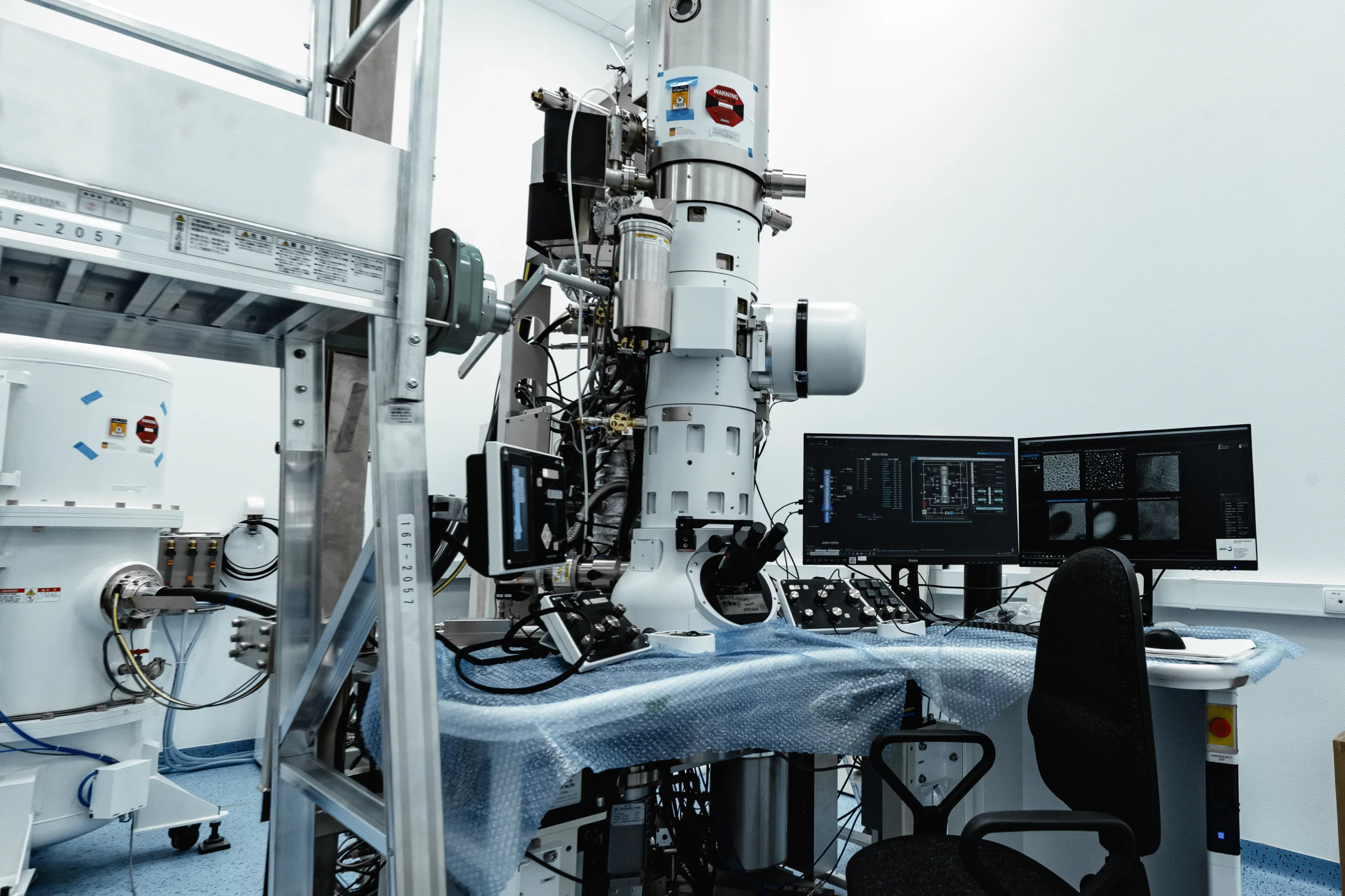
The CRYO-TEM allows the direct imaging and characterisation of objects in their natural state and aqueous solutions after they have been made glassy by shock freezing (vitrification) to prevent object distortion. The device is suitable for use at room temperature with normal material samples and with frozen preparations.
The microscope allows observation in classical parallel beam mode with a point-to-point resolution of at least 0.30 nm and in scanning focused beam mode (STEM) with a resolution of at least 0.20 nm. Image capture in TEM mode is performed using a CMOS-type camera with a resolution of at least 4kx4k pixels. The camera has a speed of 40 fps at full resolution.
The microscope is equipped with an objective lens pole piece of more than 10mm for adequate contrast of soft matter samples and for many types of microscope mounts. The STEM mode of material samples has 3 detectors. Recording of STEM images is possible at a maximum resolution of 4kx4k pixels, with automatic drift correction and on all detectors simultaneously. In the STEM mode, it is possible to analyse samples as thick as 500 nm and to obtain a large depth of field due to the low convergence angle (<1 mrad).
Equipped with a Schottky emitter electron gun integrated with a confocal Raman microscope analytical system including EDS and WDS spectrometers (SEM-EDX-WDX-Raman).
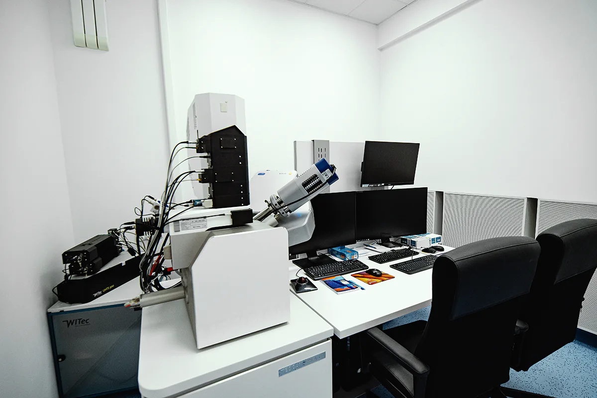
The scanning electron microscope enables correlated microscopic visualisation with simultaneous recording of Raman scattering radiation in micro areas and secondary X-ray radiation of varying wavelengths (WDX).
The resolution of SEM imaging is:
- 9nm at 15kV,
- 7nm at 1kV,
- 0nm at 500V,
- 9nm at 30kV (STEM detector).
The microscope allows electron imaging with beam tilting. High-resolution imaging of dia-, para- and ferromagnetic samples is also possible, as there are no electromagnetic fields surrounding the sample.
The primary beam can be adjusted in the accelerating voltage range from 50V to 30kV. The microscope allows simultaneous SE and BSE imaging.
Equipped with a Schottky emitter electron gun and FIB column for etching and surface modification, ion polishing system and 3D analysis software (SEM-FIB).
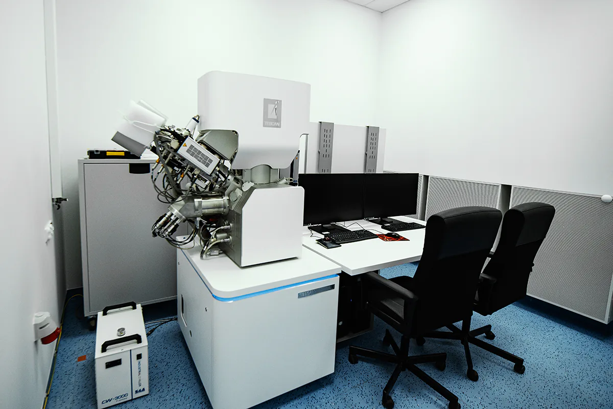
Among other things, the microscope allows imaging to sequentially capture surface-by-surface images of an object with simultaneous sectioning of several tens of nanometre-sized scraps of the preparation inside the microscope chamber during the experiment for subsequent assembly of 3D visualisations of the materials.
The SEM imaging resolution is:
- 9 nm at 15 kV,
- 7 nm at 1 kV,
- 0 nm at 500 V,
- 9 nm at 30 kV (STEM detector).
The microscope is equipped with a variable vacuum mode to compensate for the electrical charge generated during observation of non-conductive samples and with SE and BSE detectors located inside the microscope chamber and two independent detectors allowing simultaneous SE and BSE imaging.
The microscope is also equipped with an FIB (Xe) column, which is a xenon ion gun, providing the ability to rapidly modify/prepare samples.
Imaging resolution of 500 nm, ability to mount specimens with a maximum diameter of 50 cm, height of 71 cm, and weighing no more than 45 kg.
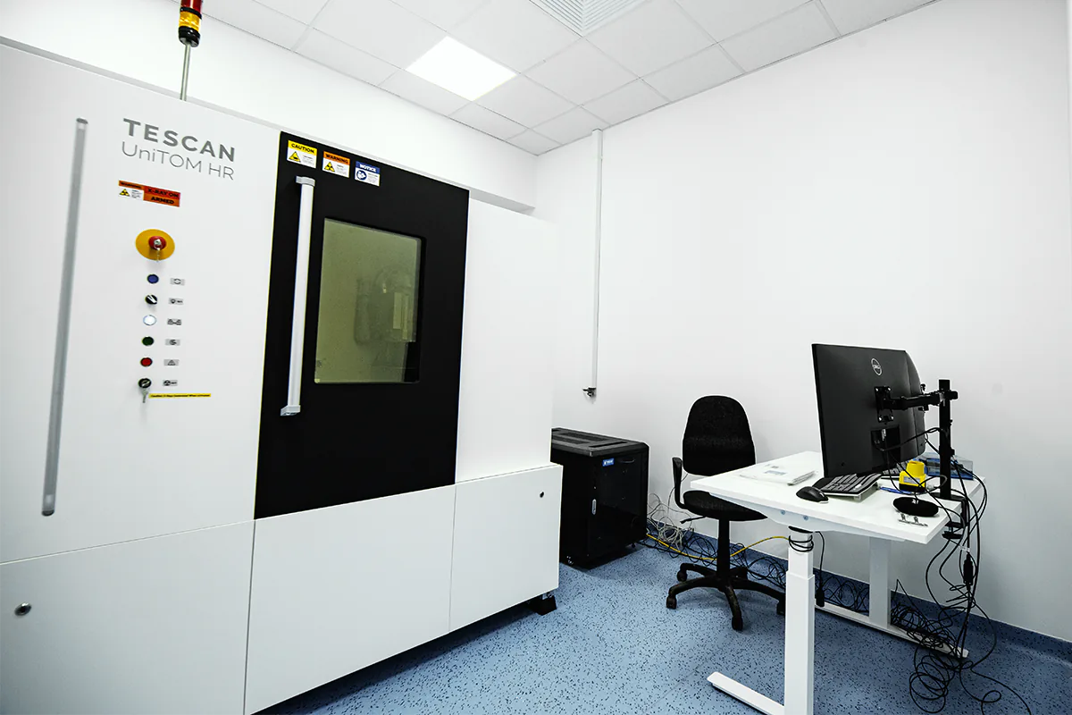
A 30-160 kV / 25W X-ray source is installed in the tomography machine. The µCt technique allows observation of dynamic processes associated with temperature change or as a result of mechanical interaction.
A high-resolution equipment featuring, among other things, a white light laser that enables surface analysis in the broad infrared range and 4D imaging.
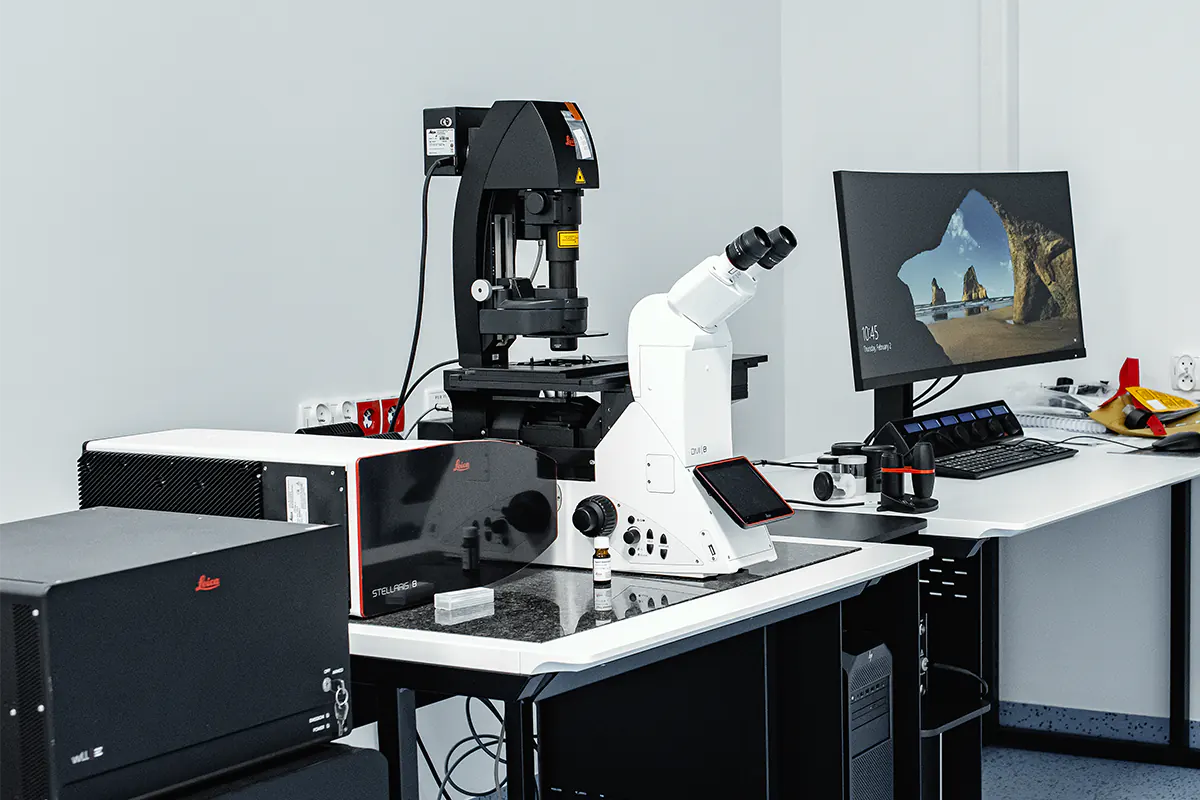
Confocal microscopy is characterised by high contrast and resolution, facilitating research in soft matter in particular. The microscope is equipped with:
- an automatic objective turret for a minimum of 6 objective lenses, with separate slider slots for Nomarski contrast (DIC), maintaining a constant distance between the objective and specimen;
- specialised objectives for confocal imaging or super-resolution (2.5x / 0.085 semi-plan apochromat, 10x/0.45 plan apochromat, 20x/0.80 plan apochromat, 40x/1.30 semi-plan apochromat, 63x /1.20 plan apochromat, 63x /1.40 plan apochromat, 100x /1.46 plan apochromat);
- a set of lasers and controls to ensure independent operation with all available laser lines (405 nm/min. 14 mW, 445 nm/min. 7.5 mW, 488 nm/min. 10 mW, 514 nm/min. 10 mW, 561 nm/min. 10 mW, 639 /min. 7.5 mW).


