

15 October
EUROPEAN BREAST CANCER DAY

„Save the date” is a series of articles that have been written to celebrate various unusual holidays. The authors of the presented materials are students, doctoral students and employees of the Faculty of Science and Technology of the University of Silesia.
15th October is celebrated as European Breast Cancer Day.
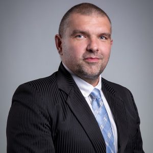
October is Breast Cancer Awareness Month, also known as the “Pink October”, initiated in 1985. Even then, people knew the importance of prevention and oncological self-awareness. We know well that in the case of diseases, especially malignant diseases, time is extremely important, and early detection of abnormalities allows for complete recovery. Armand Cholewka, PhD, DSc, MD, Associate Professor from the Institute of Biomedical Engineering speaks about methods of diagnosing breast cancer.
fot. UŚ archive
Armand Cholewka, PhD, DSc, MD, Associate Professor
Institute of Biomedical Engineering
One of the most common causes of death in women from malignancy is breast cancer. The reason for this is too late detection of existing lesions. Therefore, the incidence and the proportional mortality from breast cancer have been showing a very high growth rate for many years. In addition, standard imaging uses ionising radiation, which is known to not remain indifferent for human health. This situation shows an urgent need to develop new methods of early breast cancer detection that are quick to implement and non-invasive/completely safe and that would improve the detection of breast cancer, especially in its earliest stages of development. Under certain assumptions, thermography is one of such methods.
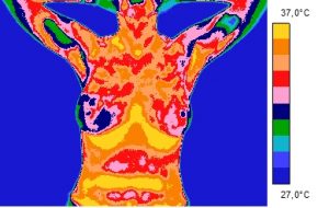
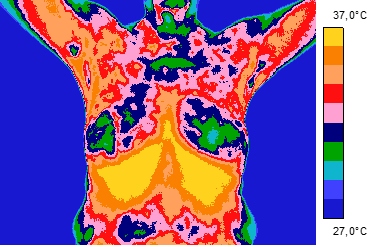
The idea of measuring the temperature of the breast gland in order to detect neoplastic foci is to make several thermograms in different projections – the front view of both breasts is shown in the following thermal images of two patients, with visible variety of colours on both breasts. Due to the fact that the colour on the heat map determines the appropriate temperature value, it can be assumed that in this case each of the visible breasts has a different temperature, i.e. there is thermal asymmetry. In turn, thermal asymmetry, i.e. the difference in temperature between the left and right breasts, may indicate the presence of neoplastic changes. The importance and usefulness of thermal imaging diagnostics has been confirmed primarily in the case of neoplastic skin lesions located on the surface and close to the body surface.
It has been proven in many studies that most neoplastic lesions were characterised by a temperature 1 – 2 degrees Celsius higher than that of a healthy breast.
When writing about the use of thermography in breast diseases, it is worth emphasising that this technique gives us information about changes in breast physiology – the course of neoplasm of blood vessels (angiogenesis) and changes in metabolic activity, and not about the structure of the gland, which is very valuable and supplementary information for methods such as mammography and sonomammography, as it allows the visualisation of physiological changes that occur long before structural changes occur.
Many of the studies have shown that patients for whom the thermogram showed thermal asymmetry of the glands, and the mammography did not show any changes, should be carried out with particular care and diagnostics performed more often. Moreover, an abnormal/highly asymmetric thermal image is assessed as ten times more important in the diagnosis of breast cancer than in the case of cancer in the patient’s family.
It should be emphasised that thermal imaging diagnostics is a quick, non-contact method that excludes exposure to harmful radiation and any discomfort of the patient. It can be used as a screening method or as an adjunct to mammography and sonomammography.
Research on the use of thermal imaging diagnostics in breast diseases and the assessment of the effects of breast cancer treatment with radiotherapy is carried out by a medical physics group from the Institute of Biomedical Engineering under the supervision of Armand Cholewka, PhD, DSc, Assoc. Prof. in cooperation with the National Institute of Oncology in Gliwice and the Medical University of Silesia.
Women, Girls – examine yourself!
Men – don’t be afraid to talk about it!