


| Agnieszka Sikora |
Scanning electron microscopy is a kind of electron microscopy which uses a much shorter light beam than the wavelength of visible light, that is, a beam of electrons, in order to study the structure of a material’s surface as well as its chemical composition and is thus able to provide a significantly better resolution than light microscopy (optical microscopy). A electron microscope is used to observe and to analyze various objects – organic and inorganic matter – from the micrometric to the nanometric scales.
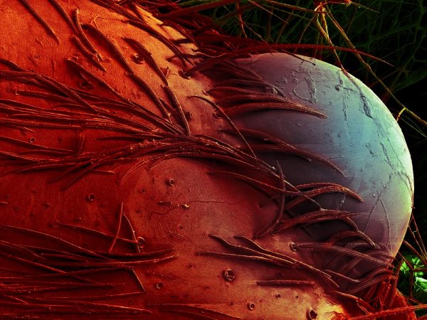
Fragment of a wolf spider’s head (Xerolycolosa nemoralis), magn. 300× | Photo: Jagna Karcz, Bartosz Baran – colorized
However, light microscopy, which was the first one to be developed, still remains the basic and most widely used microscopy technology. The first very simple microscopes were already built in the 16th century. At the end of the 17th and at the beginning of the 18th centuries, the first optical microscopes were constructed. Contemporary microscopes are devices with a high degree of automatization which are integrated with computers. Optical microscopy is not a relic of old times, since it has remained the starting point of an observation – in electron microscopy as well. When observing matter, a scientist’s first steps are accomplished by means of an optical microscope in order to initially evaluate the material at hand, and subsequently, after appropriate preparation, an electron microscope is resorted to. Scientists use various optical microscopes e.g. fluorescence microscopes which rely on the phenomena of fluorescence and phosphorescence. An innovative solution in light microscopy are confocal microscopes, as they use a laser light beam to scan probes. This analytical technology permits to research living or chemically preserved biological preparations and to create three-dimensional (3D) reconstructions of the studied object. Another kind of microscopy is scanning probe microscopy. This technology features devices which use a scanning probe in order to obtain images of the sample’s surface. An example are atomic force microscopes, which permit to achieve a three-dimensional surface image with a resolving power at the dimension range of a single atom. Due to the continuous improvements of microscopy technologies, scientists are able to achieve increasingly higher magnifications of the analyzed objects and a more detailed three-dimensional visualization of the structure of the matter.
In the case of electron microscopy, instead of a light beam, a beam of electrons is used. As a result of the interaction between the beam of electrons with the surface of the studied sample, different forms of energy are emitted. Every kind of the emitted energy is registered by means of special detectors, and subsequently it is converted into the sample’s image or into the X-ray spectrum. This very image is referred to as photomicrography, and in the case of electron scanning photography the term electron micrograph is used. The displayed image is a digital gray-scale image which is rendered by means of different techniques used to register signals emitted by the sample. It is important to emphasize that, with regard to its contents, photography refers to the nature of scientific research and is the most objective image creation technique available.
In electron microscopy, structures of plant and animal cells are analyzed at very large magnifications. The most modern electron transmission microscopes provide magnifications of several million. Due to a great sharpness depth and a very high resolution reaching 1 nm, electron scanning microscopes permit to produce almost three-dimensional images. At this point, it is expedient to add that scientists use photo editing software which enhances the effect of a 3D structure. Computed microtomography is another imaging technique which enables scientists to achieve very precise three-dimensional images of the studied objects, including their internal structure, and does not require special preparation of the sample. Contemporary scanning microscopes provide magnifications of up to a million, however, state-of-the-art microscopes reach significantly higher values. It is expedient to emphasize that a magnification of 100,000 or 200,000 enables us to see structures in the studied object which are a few dozen nanometers in size. In the majority of cases, such a magnification is sufficient.
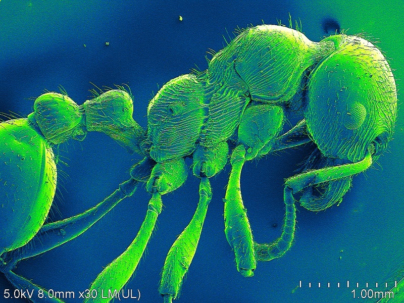
Dr. Jagna Karcz from the Institute of Biology, Biotechnology, and Environmental Protection at the Faculty of Natural Sciences of the University of Silesia is the head of the Laboratory of Electron Scanning Microscopy (SEM-Lab), and researches the development and implementation of scanning microscopy with regard to various biological objects, including the surface structure of plant epidermis exposed to environmental factors. Moreover, Dr. Karcz is one of the few specialists in carpology in this country and a popularizer of science. Since 2012, the Laboratory uses a high-resolution electron scanning microscopy with cold field emission, a cryo chamber, and an X-ray EDS spectrometer, one of the most modern microscopes of this type in Poland.
Scientists at the University of Silesia analyze various biological objects, including plant and animal tissue, bacteria, spores, plant pollen, biomaterials as well as polymers, fossil materials, and – most notably – environmental samples collected at scientific expeditions. The studied material must be appropriately prepared – a dry and conductive state is required, since in the microscopic vacuum it is prone to the destructive effects of the device’s beam of electrons. Therefore, scientists resort to many chemical procedures, which have been tested on different materials, including preservation, dehydration, drying, and sputtering, and are able to prepare the sample in such a way that the structure of the analyzed object remains intact. The biological material must be conductive, and due to this it is expedient to apply a thin layer of sputter coating consisting of a noble metal (e.g. gold). In addition, the microscope is equipped with the freezing system Cryo which permits to freeze the sample in fluid nitrogen and to observe its intact structure. The information regarding the sample’s surface structure is complemented by chemical microanalysis of the studied preparation. Due to the fact that the microscope also has an X-ray EDS spectrometer, it is possible to simultaneously obtain the image of the object’s surface and to determine its chemical composition.
An interesting example of research using SEM technology is the analysis of surface degradation processes in plastic materials caused by microorganisms. Microbiologists research, among others, biofilm bacteria, zoologists analyze insect morphologies for taxonomic purposes, and botanists inquire into environmental effects on plant populations e.g. by means of analyzing structures of invasive species in connection with molecular research.
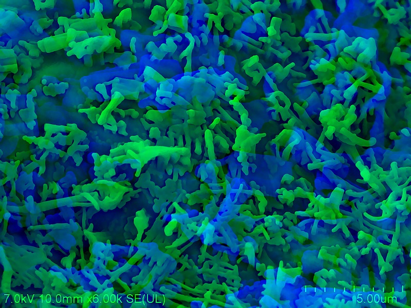
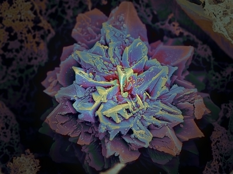
Since many years, Dr. Karcz has been a researcher and photographer of microworlds. It is expedient to add at this point that the original images produced by an electron microscope are black and white, including all shades of gray, and could be obtained due to the secondary electrons emitted by the samples. In these samples, the contrast is linked to their topography – convex parts are bright, concave fragments – dark. The contents of microphotographs refer to scientific research and to didactic courses in various academic disciplines, such as biology, biotechnology, and environmental protection. The themes of these photographs are very diverse. They depict natural surfaces of various objects, that is, how they really look like in their natural environment, as well as structures of plants and animals modified by environmental factors (biotic and abiotic). It is possible to admire the extraordinary structure of cobwebs and their complex spatial configuration. Moreover, they are a perfect material for the production of matrices which enable scientists to grow various animal cells. The photographs also document the process of degradation of plastic materials by microorganisms and the elaborate construction of sponge skeletons which are found in water bodies. In her works, Dr. Karcz demonstrates that science can be an inspiration for and theme of photography, and that the world which surrounds us is both fascinating and mysterious.
The article entitled ‘Fascinating microworlds’ was published in ‘No Limits’, no. 1 (1) in 2020.